
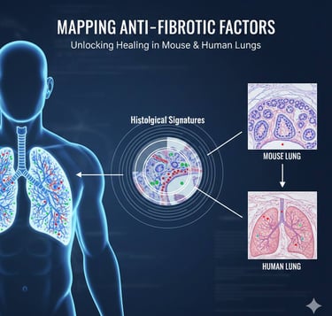
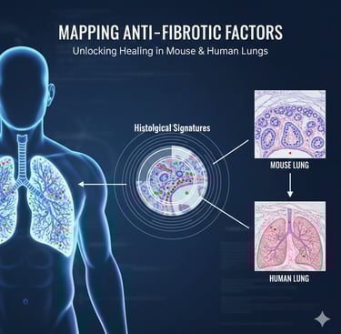
Histological signatures reveal anti-fibrotic mechanisms in human and mouse lungs
Blog post description.
1/1/20262 min read
Pulmonary fibrosis is a serious and often progressive lung disease in which normal lung tissue is gradually replaced by scar tissue. This scarring reduces lung elasticity, limits oxygen exchange, and leads to chronic shortness of breath. Despite advances in medicine, current treatments mainly slow disease progression and do not fully reverse lung damage. Understanding how healthy lung tissue naturally resists fibrosis is therefore a major goal in biomedical research.
In a recent study published in Nature, researchers explored lung tissue at the histological level to uncover natural anti-fibrotic mechanisms—biological processes that actively prevent or limit scar formation
What the Researchers Studied
The team analyzed lung samples from both humans and mouse models, comparing healthy tissue with fibrotic lungs. Instead of focusing only on molecular data, they examined how tissue structure and cellular organization reflect protective biological signals.
Their goal was to identify histological signatures—distinct tissue patterns—that correlate with factors known to suppress fibrosis
Key Findings
The study revealed that certain lung regions show consistent structural features associated with anti-fibrotic activity. These regions were enriched with signals that:
• Suppress excessive fibroblast activation
• Limit abnormal collagen deposition
• Support tissue repair rather than permanent scarring
Importantly, these protective signatures were observed in both human and mouse lungs, suggesting that core anti-fibrotic mechanisms are evolutionarily conserved.
The researchers also found that when these protective tissue patterns are lost or disrupted, the lung becomes more vulnerable to progressive fibrosis.
Why This Matters for Human Health
Pulmonary fibrosis affects millions worldwide and is often irreversible once advanced. This study shifts the perspective from asking “Why does fibrosis happen?” to “How does the lung normally prevent fibrosis?”
Understanding these natural defense mechanisms could:
• Enable earlier detection of fibrotic risk
• Help stratify patients based on disease progression
• Reveal new therapeutic targets that enhance the body’s own anti-fibrotic responses
Rather than only blocking scar formation, future treatments may aim to restore or
reinforce protective tissue programs.
Broader Implications
Fibrosis is not limited to the lungs—it also affects organs such as the liver, kidneys, and heart. The principles uncovered in this study may therefore apply to multiple chronic diseases characterized by tissue scarring.
By linking tissue structure to functional health outcomes, this research highlights the continuing importance of histology in modern medicine—not just as a diagnostic tool, but as a window into disease resistance and regeneration.
Conclusion
This Nature study provides compelling evidence that lung tissue carries built-in anti-fibrotic programs that can be identified at the histological level. Recognizing and understanding these protective patterns opens new avenues for treating pulmonary fibrosis and potentially other fibrotic diseases. It represents a shift toward therapies that work with the body’s natural defense systems rather than against irreversible damage.

Follow
Contact
info@microlab.ink
© Microlab-copyright 2026
