
Histology image analysis of 13 healthy tissues reveals molecular-histological correlations
3/13/20262 min read
Histology has traditionally focused on studying the microscopic structure of tissues. However, modern research is now combining histology with molecular biology and artificial intelligence to uncover deeper biological insights.
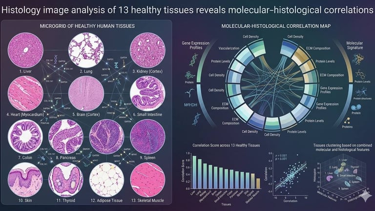
A recent study analyzed histology images from 13 different healthy human tissues and investigated how the microscopic appearance of cells relates to gene expression patterns. Researchers used high-resolution whole-slide images stained with hematoxylin and eosin (H&E) together with RNA sequencing data to explore this connection.
From Microscopic Images to Molecular Data
Each histology slide contains thousands of cells, and analyzing them manually can be extremely time-consuming.
To overcome this challenge, the researchers used a deep-learning framework that automatically identifies and segments cell nuclei in tissue sections.
By extracting quantitative features from these nuclei—such as shape, size, intensity, and spatial distribution—the system could characterize tissue morphology with high precision.
These morphological features were then compared with gene expression profiles obtained from the same tissues.
What the Study Found
The results showed a strong relationship between nuclear morphology and RNA expression patterns across different organs.
In other words, the way a cell nucleus looks under the microscope can reflect the molecular activity occurring inside the cell.
The analysis also identified specific gene sets associated with particular nuclear features, many of which are involved in:
• cell growth and development
• metabolism
• immune system functions
• intercellular communication
This suggests that histological patterns are not just structural observations—they can also provide clues about the underlying biological processes.
Why This Matters for Medicine
Traditionally, histology relies heavily on visual interpretation by pathologists. While expert evaluation is extremely valuable, it can also be subjective and time-consuming.
By integrating histology, genomics, and artificial intelligence, scientists can:
• identify new biomarkers
• improve disease diagnosis
• understand how tissues function at multiple biological levels
• detect early molecular changes before visible pathology appears
The Future of Histology
This research highlights the emerging field of imaging genomics, where microscopic tissue images are analyzed together with molecular data.
In the future, combining histology with advanced computational tools could transform pathology from a primarily visual discipline into a quantitative and predictive science, providing deeper insights into human health and disease.

Follow
Contact
info@microlab.ink
© Microlab-copyright 2026
