
Mapping the landscape of histomorphological cancer phenotypes using self-supervised learning on unannotated pathology slides
1/29/20262 min read
The Study: AI Meets Cancer Histology
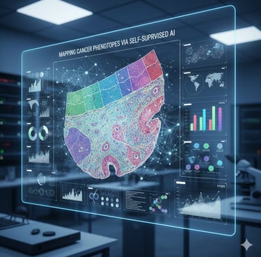
In a groundbreaking study published in Nature Communications, researchers applied self-supervised deep learning to analyze whole-slide images of lung adenocarcinoma, a common type of lung cancer. Unlike traditional methods that rely on manual labeling by pathologists, this AI-driven approach learns patterns directly from the images themselves.
The algorithm scanned thousands of histology slides and automatically identified distinct tissue and cellular structures, such as tumor cells, supportive stromal tissue, and immune cell infiltration areas. These structures, or “histomorphological phenotypes,” reveal the diversity and complexity of tumors in ways that are often impossible to quantify by the human eye alone.
Key Findings
The study demonstrated several important points:
1. Hidden prognostic signals: The AI discovered tissue patterns that strongly correlated with patient survival.
This means that histology images contain more prognostic information than previously realized.
2. Tumor heterogeneity revealed: By distinguishing subtle differences in tissue architecture, the model highlighted the diverse environments within tumors, including areas of immune response, fibrosis, and tumor growth.
3. Bridging morphology and molecular data: Researchers connected histological features to molecular and clinical data, providing a richer understanding of how tumor structure relates to genetics, biology, and outcomes.
Why This Matters
Histology has been a cornerstone of cancer diagnosis for over a century, but it is inherently limited by what humans can see. By combining histology with machine learning, we can now:
• Identify patterns that predict how aggressive a tumor is.
• Understand how tumors interact with the immune system and surrounding tissues.
• Discover potential targets for therapy based on tissue architecture.
In other words, AI doesn’t replace pathologists—it enhances their vision, helping clinicians and researchers uncover hidden insights in the microscopic world of cancer.
Conclusion
This study represents a significant step toward precision oncology, where treatment and prognosis are informed not just by genetic data but also by detailed tissue morphology. By harnessing AI to read histology slides, researchers are unlocking a new layer of information that could improve cancer diagnosis, treatment planning, and patient outcomes.
The microscopic patterns in tumors are no longer just beautiful images under the microscope—they are valuable data waiting to be decoded.

Follow
Contact
info@microlab.ink
© Microlab-copyright 2026
