
Revealing 3D microanatomical structures of unlabeled thick cancer tissues using holotomography and virtual H&E staining
2/19/20261 min read
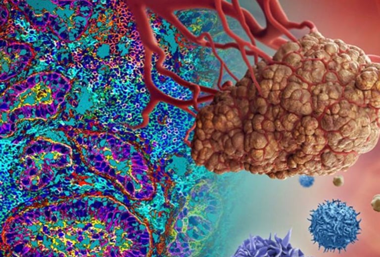
Imagine being able to see cancer tissues in full 3D without slicing them at all — not just a few 2D sections under a microscope. This is exactly what researchers accomplished in this groundbreaking study published in Nature Communications.
What They Did
Traditionally, histopathology (the gold standard for diagnosing diseases like cancer) relies on thin 2D slices of tissue stained with Hematoxylin & Eosin (H&E). But this limits us to flat images and loses crucial 3D context — such as how cells are arranged deep inside tumors.
To overcome this, the researchers:
1. Captured the tissue’s 3D refractive index (RI) using a technique called holotomography — a label‑free optical method that maps internal structures without staining.
2. Trained a deep learning model to translate that RI data into virtual H&E images in 3D. These images mimic traditional stained microscopy but preserve spatial relationships and depth information.
3. Applied this framework to thick biopsy samples (up to 50 µm — far thicker than conventional slides) from colon and gastric cancer tissues.
Why It Matters
• Preserves 3D structure: Instead of reconstructing multiple slices, the method reveals
microanatomical details like glands, lumens, and cell nuclei throughout the entire volume.
• Subcellular resolution: The results show detailed organization within tissues, not just on a surface.
• Valid and scalable: The virtual H&E results were compared with traditional staining to ensure accuracy and reproducibility across different samples and institutions.
In Simple Terms
This research brings histology into the third dimension. By combining optical imaging and AI‑driven virtual staining, scientists can now visualize how cells and structures actually exist in space — a major leap forward in diagnostic imaging and cancer research.
Source link: https://www.nature.com/articles/s41467-025-59820-0
Follow
Contact
info@microlab.ink
© Microlab-copyright 2026
