Introduction
Identifying histology slides under time pressure can be one of the most challenging aspects for medical students during exams. With the clock ticking, mastering slide IDs can make all the difference in your performance. This post will share effective strategies to help you quickly recognise key histological features, ensuring you make the most of your exam time.
Why It Matters in Exams
Histology slide identification is a critical component of many medical exams. The ability to swiftly and accurately identify slides can significantly influence your overall score. Teachers often focus on common tissues and pathological changes, so knowing what to look for can help you excel in histology, pathology, and even embryology sections of your exams.
Common Mistakes to Avoid
- Overlooking Key Features: Students often miss crucial identifying characteristics, such as cell shape, arrangement, or staining properties.
- Rushing Through Slides: In the heat of the moment, rushing can lead to misidentification; take a breath and assess systematically.
- Ignoring Context: Understanding the tissue’s location and function can provide vital clues for identification.
Strategies for Success
Here are some practical strategies to help you identify histology slides quickly and accurately:
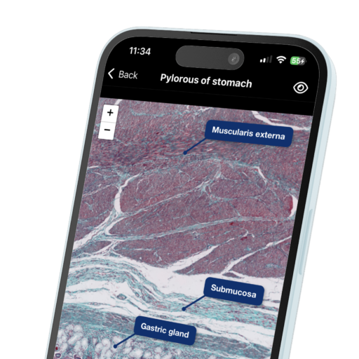
- Familiarise Yourself with Common Tissues: Start with the basics. Familiarise yourself with the appearance of common tissues such as epithelium, connective tissue, muscle, and nervous tissue. Use diagrams and images to visualise different types and their characteristics.
- Master the Stains: Different stains highlight various structures; know what each stain emphasises. For example, H&E staining gives contrasting colours to nuclei and cytoplasm, while special stains (like Masson’s trichrome) can help identify connective tissues.
- Use Mnemonics: Create mnemonics to remember specific features of tissues. For instance, for identifying cardiac muscle, remember “C for Cross-striations” and “A for Intercalated discs.”
- Practice with Timed Quizzes: Simulate exam conditions by using flashcards or online resources that require you to identify slides under timed conditions. This will help increase your speed and confidence.
- Group Study Sessions: Collaborate with peers to quiz each other on slide identification. Teaching others can reinforce your own knowledge.
How to Remember It
To help with quick identification, follow this simple step-by-step approach:
- Initial Scan: Quickly scan the slide to get an overall impression of the tissue type.
- Identify Key Features: Look for distinctive characteristics, like cell shape (squamous, cuboidal, columnar), arrangement (simple, stratified, pseudostratified), and any special structures (like cilia or goblet cells).
- Match with Known Tissues: Compare the identified features against your mental library of common tissues.
- Consider the Function: Think about the organ or system the tissue belongs to, which can provide context clues for identification.
Conclusion
As you prepare for your histology exams, remember that practice and familiarity are key. Implement these strategies and take the time to build your knowledge of histological features. With organised study and effective techniques, you’ll be well-equipped to handle slide identification under pressure.
For additional support, practice with interactive flashcards and quizzes on Microlab.


Leave a Reply