Identifying Epithelial Tissue Types in Histology
As a first-year medical student, mastering the identification of epithelial tissue types under Haematoxylin and Eosin (H&E) staining is crucial. Epithelial tissues serve as protective barriers, are involved in absorption, secretion, and sensation, and understanding their types can greatly enhance your histological skills and exam performance.
Why It Matters in Exams
In histology exams, you may be presented with various slides of tissues, and identifying epithelial types is a common question. Questions may ask you to describe the function, location, and characteristics of different epithelia based on microscopy images. This knowledge not only helps in histology but also links with anatomy and physiology, making it a multidisciplinary topic. Recognising these tissues can also assist in understanding pathological conditions, as many diseases affect epithelial integrity.
Types of Epithelial Tissue
Epithelial tissues are classified based on the number of layers and the shape of the cells:
- Simple Epithelium: A single layer of cells.
- Stratified Epithelium: Multiple layers of cells.
Cell shapes include:
- Squamous: Flat and thin.
- Cuboidal: Cube-shaped.
- Columnar: Taller than they are wide.
Examples of epithelial types include:
- Simple Squamous Epithelium: Found in alveoli of lungs and lining blood vessels.
- Cuboidal Epithelium: Common in kidney tubules.
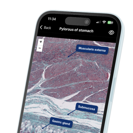
- Simple Columnar Epithelium: Lines the gastrointestinal tract.
- Stratified Squamous Epithelium: Found in the skin and lining of the mouth.
How to Remember It
To help remember the different types of epithelial tissues, you can use a simple mnemonic:
SCC – Simple Cuboidal Columnar
This can be expanded to include the shapes:
SSCC – Simple Squamous, Cuboidal, Columnar
Additionally, visualising where these tissues are found can aid in memorisation. Create flashcards with images of the epithelial types on one side and their functions and locations on the other.
Tips for Slide Reading
- Start with the basics: Look for the number of layers first. This will help you narrow down your options.
- Identify cell shape: Once you know the layers, examine the shape of the cells to determine the specific type.
- Watch for special features: Look for cilia or keratinisation, as these can give clues to the function of the tissue.
- Practice, practice, practice: Use resources like histology atlases and online galleries to familiarise yourself with various slides.
By mastering these skills, you’ll not only excel in your histology exams but also build a solid foundation for your future studies in medicine.
Practice with interactive flashcards and quizzes on Microlab to enhance your understanding of histology and improve your exam readiness!


Leave a Reply